
Abstract
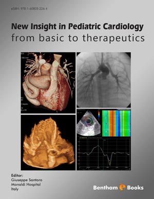
Magnetic Resonance Imaging (MRI) has emerged as a valuable non-invasive diagnostic tool in congenital heart malformations providing anatomical and functional data regardless of patient’s size and quality of thoracic window. This technique is particularly indicated to avoid cardiac catheterization in post-surgical adult patients in whom the echocardiographic window is often poor. MRI is able to provide both accurate 3-dimensional images of the cardiovascular system as well as precisely quantify volumes and mass of the cardiac chambers and functional data of any single segment of the heart. MRI is now considered a Class I indication in pediatric or adult patients with congenital heart malformations. It is able to define morphological data of cardiac malformation and its functional consequences at the same time. This paper summarizes the most relevant technical aspects of MRI in congenital heart disease and reports on useful protocols to evaluate the most common malformations.
Keywords: Magnetic resonance imaging, congenital heart disease, morphological assessment, right ventricle, ventricular function, aortic coarctation, pulmonary branch stenosis, septal defect, single ventricle, tetralogy of Fallot.
Related Journals

Anti-Inflammatory & Anti-Allergy Agents in Medicinal Chemistry

Current Diabetes Reviews

Current Neurovascular Research

Current Respiratory Medicine Reviews

Current Pediatric Reviews

Infectious Disorders - Drug Targets

Current Stem Cell Research & Therapy

Endocrine, Metabolic & Immune Disorders - Drug Targets
Current Alzheimer Research

Current Aging Science
Related Books

Andrology: Current and Future Developments
Male Infertility: An Integrative Manual of Western and Chinese Medicine

Awake Thoracic Surgery
The Anatomical Foundations of Regional Anesthesia and Acute Pain Medicine Macroanatomy Microanatomy Sonoanatomy Functional anatomy
Frontiers in Anti-infective Agents
Frontiers in Anti-infective Agents

Frontiers in Anti-infective Agents

Frontiers in Anti-infective Agents

Frontiers in Anti-infective Agents

Frontiers in Anti-infective Agents