Generic placeholder image

Current Rheumatology Reviews

Editor-in-Chief

ISSN (Print): 1573-3971
ISSN (Online): 1875-6360

Case Report

Late Presentation of Primary Oxalosis, Microcrystalline Arthropathy, and Tumoral Calcinosis: A Case Report and a Literature Review

Author(s): Sanda Mrabet* and Mohamed BenHmida

Volume 20, Issue 4, 2024

Published on: 15 January, 2024

Page: [455 - 458] Pages: 4

DOI: 10.2174/0115733971271874231118154332

Price: $65

Abstract

Background: Primary hyperoxaluria consists of a group of inherited disorders with enzymatic defects in the glyoxylate pathway, leading to decreased oxalate metabolism. The resulting oxalic deposition is specifically responsible for kidney disease and joint disease. Neonatal oxalosis is the most severe form of primary hyperoxia type 1, with the onset of end-stage renal disease in childhood.

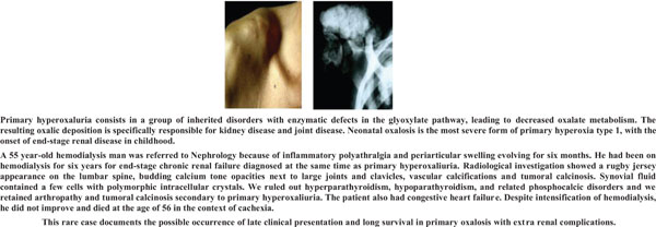
Case Presentation: A 55-year-old hemodialysis man was referred to Nephrology because of inflammatory polyarthralgia and periarticular swelling evolving for six months. He had been on hemodialysis for six years for end-stage chronic renal failure, diagnosed at the same time as primary hyperoxaluria. Radiological investigation showed a rugby jersey appearance on the lumbar spine, budding calcium tone opacities next to large joints and clavicles, vascular calcifications and tumoral calcinosis. The synovial fluid contained a few cells with polymorphic intracellular crystals. We ruled out hyperparathyroidism, hypoparathyroidism, and related phosphocalcic disorders, and we retained arthropathy and tumoral calcinosis secondary to primary hyperoxaliuria. The patient also had congestive heart failure. Despite intensification of hemodialysis, he did not improve and died at the age of 56 in the context of cachexia.

Conclusion: This rare case documents the possible occurrence of late clinical presentation and long survival in primary oxalosis with extra renal complications.

Keywords: Primary hyperoxaluria, calcinosis, dialysis, arthritis, synovial fluid, phosphocalcic disorders, hemodialysis.

Graphical Abstract
[1]
Lorenz EC, Michet CJ, Milliner DS, Lieske JC. Update on oxalate crystal disease. Curr Rheumatol Rep 2013; 15(7): 340.
[http://dx.doi.org/10.1007/s11926-013-0340-4] [PMID: 23666469]
[2]
van Woerden CS, Groothoff JW, Wanders RJ, Davin JC, Wijburg FA. Primary hyperoxaluria type 1 in The Netherlands: Prevalence and outcome. Nephrol Dial Transplant 2003; 18(2): 273-9.
[http://dx.doi.org/10.1093/ndt/18.2.273] [PMID: 12543880]
[3]
M’dimegh S, Aquaviva-bourdain C, Omezzine A, et al. HOGA1 gene mutations of primary hyperoxaluria type 3 in tunisian patients. J Clin Lab Anal 2017; 31(3): e22053.
[http://dx.doi.org/10.1002/jcla.22053] [PMID: 27561601]
[4]
Nagara M, Tiar A, Ben Halim N, et al. Mutation spectrum of primary hyperoxaluria type 1 in Tunisia: Implication for diagnosis in North Africa. Gene 2013; 527(1): 316-20.
[http://dx.doi.org/10.1016/j.gene.2013.06.023] [PMID: 23810941]
[5]
Horta-Baas G, Vargas-Gutiérrez C, Barile-Fabris L. Large joint destructive arthropathy and tumoral calcinosis associated to primary oxalosis: Case report and literature review. Reumatol Clin 2013; 9(3): 181-5.
[http://dx.doi.org/10.1016/j.reumae.2013.04.002]
[6]
Lorenzo V, Torres A, Salido E. Primary hyperoxaluria. Nefrologia 2014; 34(3): 398-412.
[http://dx.doi.org/10.3265/Nefrologia.pre2014.Jan.12335]
[7]
Fisher D, Hiller N, Drukker A. Oxalosis of bone: Report of four cases and a new radiological staging. Pediatr Radiol 1995; 25(4): 293-5.
[http://dx.doi.org/10.1007/BF02011105] [PMID: 7567242]
[8]
Bakshi NA, Al-Zahrani H. Bone marrow oxalosis. Blood 2012; 120(1): 8.
[http://dx.doi.org/10.1182/blood-2011-12-400192] [PMID: 22953330]
[9]
Strauss SB, Waltuch T, Bivin W, Kaskel F, Levin TL. Primary hyperoxaluria: Spectrum of clinical and imaging findings. Pediatr Radiol 2017; 47(1): 96-103.
[http://dx.doi.org/10.1007/s00247-016-3723-7] [PMID: 27844104]
[10]
Nachite F, Dref M, Fakhri A, Rais H. Primary hyperoxaluria detected by bone marrow biopsy: Case report. BMC Clin Pathol 2017; 17(1): 20.
[http://dx.doi.org/10.1186/s12907-017-0059-7] [PMID: 28943803]
[11]
Cochat P, Rumsby G. Primary hyperoxaluria. N Engl J Med 2013; 369(7): 649-58.
[http://dx.doi.org/10.1056/NEJMra1301564] [PMID: 23944302]
[12]
El Hage S, Ghanem I, Baradhi A, et al. Skeletal features of primary hyperoxaluria type 1, revisited. J Child Orthop 2008; 2(3): 205-10.
[http://dx.doi.org/10.1007/s11832-008-0082-4] [PMID: 19308578]
[13]
Zhou H, Yang M, Zou Y. Uraemic tumoral calcinosis in a peritoneal dialysis patient. Intern Med J 2018; 48(12): 1544-6.
[http://dx.doi.org/10.1111/imj.14130] [PMID: 30517983]
[14]
Yano H, Kinjo M. Tumoral calcinosis. Cleve Clin J Med 2021; 88(4): 208-9.
[http://dx.doi.org/10.3949/ccjm.88a.20084] [PMID: 33795242]
[15]
Marangella M, Petrarulo M, Cosseddu D, Vitale C, Linari F. Oxalate balance studies in patients on hemodialysis for type I primary hyperoxaluria. Am J Kidney Dis 1992; 19(6): 546-53.
[http://dx.doi.org/10.1016/S0272-6386(12)80833-X] [PMID: 1595703]
[16]
Verbruggen LA, Bourgain C, Verbeelen D. Late presentation and microcrystalline arthropathy in primary hyperoxaluria. Clin Exp Rheumatol 1989; 7(6): 631-3.
[PMID: 2612083]
[17]
Mrabet S, Chaabouni Y, Hmida MB. Acute kidney injury leading to the diagnosis of sporadic testicular Burkitt’s lymphoma: A case report. Int J Surg Case Rep 2022; 97: 107471.
[http://dx.doi.org/10.1016/j.ijscr.2022.107471] [PMID: 35933951]

Rights & Permissions Print Cite
© 2024 Bentham Science Publishers | Privacy Policy